Brain Tumor Classification
Classifying Brain Tumor using Advanced Machine Learning
Brain Tumor Classification
Classify tumor types using AI models
Project Abstract
Understanding our approach to brain tumor classification
Brain tumors are among the most serious medical conditions that require early and accurate diagnosis for effective treatment. This project presents an advanced deep learning approach for automated brain tumor detection using MRI scan analysis.
Our system employs a Convolutional Neural Network (CNN) to perform binary classification, determining whether a brain tumor is present or absent in MRI scans. The model was trained on a comprehensive dataset of labeled MRI images with "Yes" and "No" classifications for tumor presence.
The implementation achieved an impressive 97.03% training accuracy and 92.16% validation accuracy, demonstrating the effectiveness of our CNN architecture in medical image analysis. The model processes 100x100 pixel MRI images and provides confident predictions with probability scores.
Tumor Categories
Our AI system can detect the presence of brain tumors in MRI scans
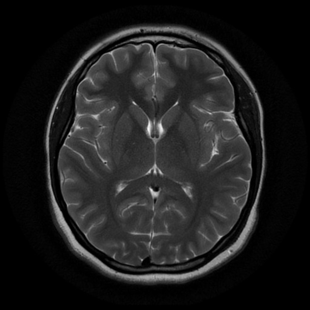
Normal brain tissue without any tumor presence. Healthy brain scans showing no abnormal growths or masses.
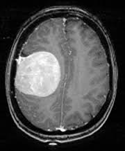
Brain scan showing presence of tumor tissue. Requires immediate medical attention and further analysis.
Interactive Prediction
Upload an MRI scan to classify brain tumor type
Drag & drop an MRI image
or click to select (JPEG, PNG, BMP, TIFF)
Upload an image to see classification results
Our CNN Model Statistics
Performance metrics and training results
97.03%
Final Accuracy
Training Accuracy
92.16%
Validation Accuracy
Cross-validation Score
0.0855
Final Loss
Training Loss
0.3086
Validation Loss
Validation Loss
Data Preprocessing & Model Building
Our approach to building an effective brain tumor detection system
Collected MRI brain scan images with binary labels (Yes/No for tumor presence)
- Total Images: 200+ MRI scans
- Tumor Present: 100 images
- No Tumor: 100 images
- Image Format: JPEG, PNG
- Resolution: 100 x 100 pixels
Applied various preprocessing techniques to enhance image quality and normalize data
- Image Resizing: 100 x 100 pixels
- Normalization: Pixel values [0,1]
- Data Augmentation: Rotation, Flip, Zoom
- Train/Validation Split: 80/20
- Batch Size: 32
Built a Sequential CNN model optimized for binary classification
- Model Type: Sequential CNN
- Total Parameters: 4,353,346
- Model Size: 16.61 MB
- Activation: ReLU, Softmax
- Optimizer: Adam
Achieved excellent performance with high accuracy and low loss
- Final Accuracy: 97.03%
- Validation Accuracy: 92.16%
- Final Loss: 0.0855
- Validation Loss: 0.3086
- Training Epochs: 10

Training logs showing progressive improvement in accuracy and reduction in loss over 10 epochs

Convolutional Layers
- • Conv2D: 32 filters, 3x3 kernel
- • MaxPooling2D: 2x2 pool size
- • Conv2D: 64 filters, 3x3 kernel
- • MaxPooling2D: 2x2 pool size
Dense Layers
- • Flatten Layer
- • Dense: 128 neurons, ReLU
- • Dropout: 0.5 rate
- • Dense: 2 neurons, Sigmoid
Model Summary
- • Total Parameters: 4,353,346
- • Trainable Parameters: 4,353,346
- • Model Size: 16.61 MB
- • Input Shape: (224, 224, 3)